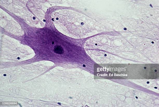
motor neuron; spinal cord, 50x at 35mm. shows: cell body, nucleus, dendrites (numerous processes attached to cell body), axon (single, long, nerve fiber), and neuroglial cells (dark spots). - motor neuron stock-fotos und bilder
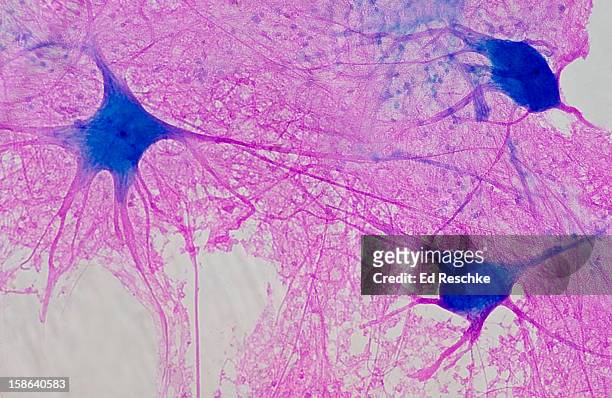
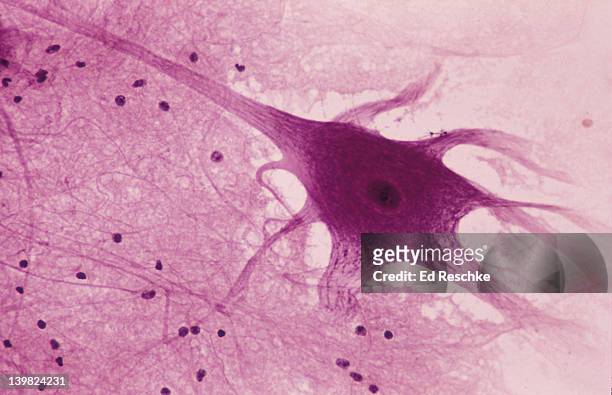
motor neuron (multipolar) with cell body and many processes (mostly dendrites), spinal cord (magnification x100). this multipolar motor neuron comes from the anterior (ventral) horn of the spinal cord grey matter. the stain is methylene blue and phloxine. - motor neuron stock-fotos und bilder
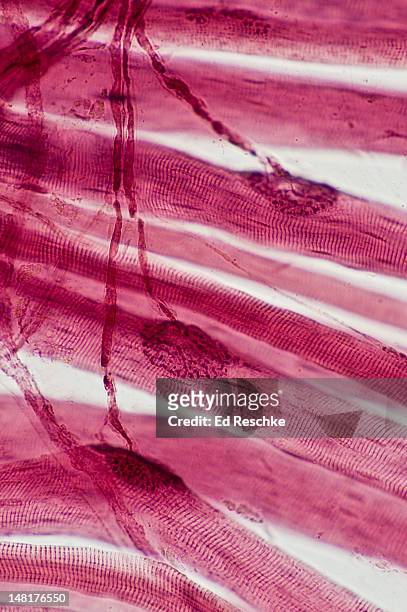
neuromuscular junctions, motor neurons, skeletal muscle fibers (cells) 25x at 35mm - motor neuron stock-fotos und bilder
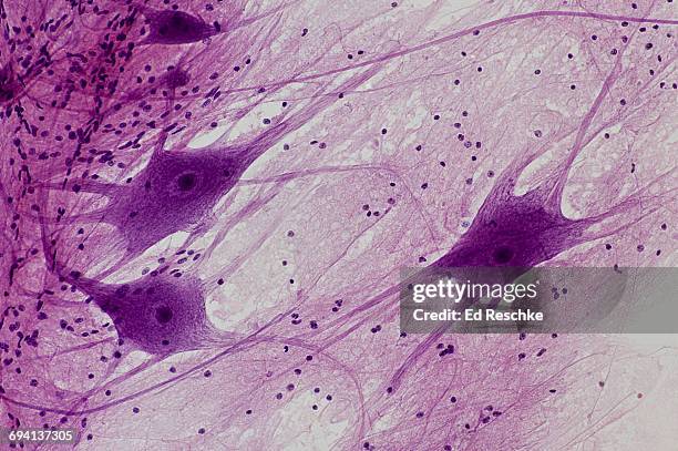
neurons (motor), spinal cord, 50x at 35mm. shows: 3 neurons, cell bodies, nuclei, dendrites, probable axon, and neuroglial cells. - motor neuron stock-fotos und bilder
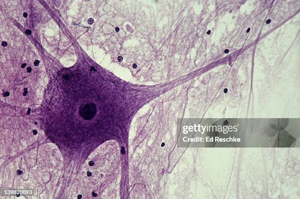
neuron (motor), spinal cord, 100x at 35mm. shows: cell body, nucleus, dendrites (several), axon (single, long nerve fiber), and neuroglial cells (black spots). - motor neuron stock-fotos und bilder
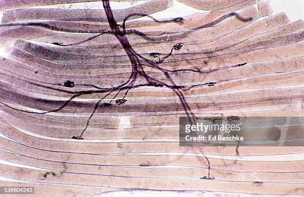
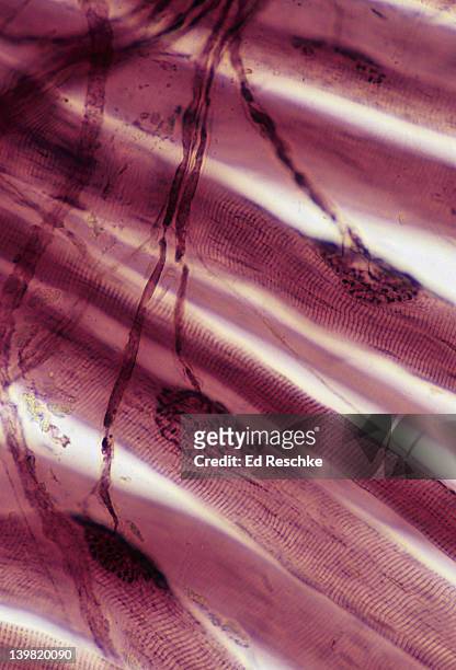
neuromuscular junctions, motor neurons and skeletal muscle fibres (cells). (magnification x100). showing two neuromuscular junctions, motor neuron axons and striations in the skeletal muscle fibres. - motor neuron stock-fotos und bilder
motor neuron; spinal cord, 50x at 35mm. shows: cell body, nucleus, dendrites (numerous processes attached to cell body), axon (single, long, nerve fiber), and neuroglial cells (dark spots). - motor neuron stock-fotos und bilder
photomicrograph of spinal cord neuron of the grey matter showing cell body, nucleus and neucleolus; 250x. - motor neuron stock-fotos und bilder
neuron (motor), spinal cord, 50x at 35mm. shows: cell body, nucleus, dendrites (several), axon (single, long nerve fiber), and neuroglial cells (black spots). - motor neuron stock-fotos und bilder
photomicrograph of motor neuron of spinal cord, showing cell body, nucleus, dendrites, axon, and neurolglia (black spots); 50x. - motor neuron stock-fotos und bilder
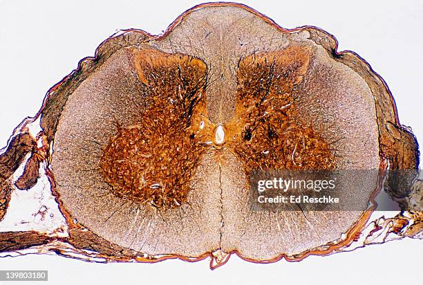
spinal cord. cross section, 5x shows: gray matter (inner pink, butterfly-shaped area), white matter (outer blue area), central canal, meninges, dorsal horn, lateral horn, ventral (anterior) horn, and anterior horn cells (motor neuron cell bodies). - motor neuron stock-fotos und bilder
Motor neuron synapsing with skeletan muscle cells. Brain exhibition Inside MIT Museum Building at 265 Massachusetts Avenue Cambridge, Boston...
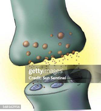
Motor End Plates. Motor End Plates Are The Neuromuscular Junctions That Conduct The Nerve Impulse Action Potential From The Axon To The Muscle Fiber.
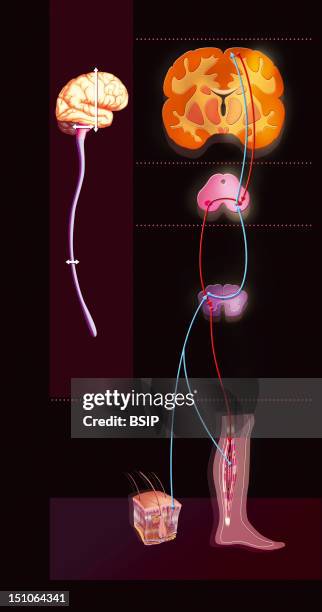
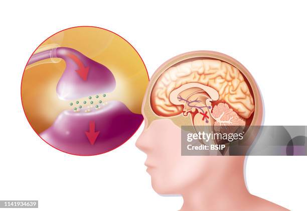
Sensorimotor Loop. Representation Of The Sensorimotor Loop Monitoring Of The Brain To Link The Stimuli Originating From The Sensorial Receptors Par...
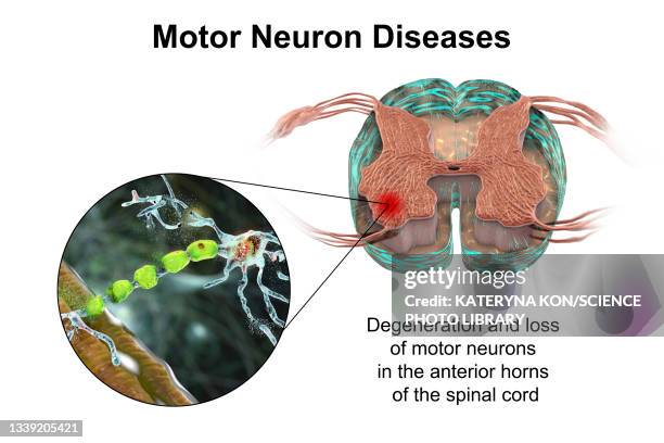
degradation of motor neurons, illustration - motor neuron stock-grafiken, -clipart, -cartoons und -symbole
Motor End Plate Or Neuromuscular Synapse Junction Between The Efferent Neuron And The Muscle Fiber. Optic Microscopy About X 150 The Size Of An Image...
Sensorimotor Loop. Representation Of The Sensorimotor Loop Monitoring Of The Brain To Link The Stimuli Originating From The Sensorial Receptors Par...
polio viruses affecting motor neurons, illustration - motor neuron stock-grafiken, -clipart, -cartoons und -symbole
neuron (motor), spinal cord, 50x at 35mm. shows: cell body, nucleus, dendrites (several), axon (single, long nerve fiber), and neuroglial cells (black spots). - motor neuron stock-fotos und bilder
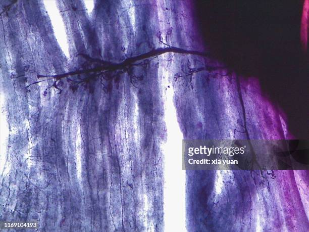
neuromuscular junction. 250x at 35mm. shows: a neuromuscular junction (motor end plate), an axon of a motor neuron, and a striated skeletal muscle fiber (cell). - motor neuron stock-fotos und bilder
Illustration of Parkinson's disease. It affects dopamine neurons in the substantia nigra. Dopamine, vital for transmitting the nerve impulse used in...
Illustration of a synapse between two dopamine neurons with Parkinson's disease, showing a decrease in nerve impulse transmission, due to a decrease...
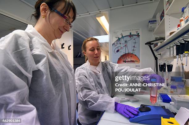
Marie-Reine Schneider, head of the pharmaceutical development and Caroline Gouarne, researcher in cell biology, use a solution of a molecule called...
Photo Essay From Laboratory. Genetics Research Laboratory. Gene Therapy Requires A Gene Drug And A Vector To Carry It To The Target Cell. The Study...
Genetics Research Laboratory. Gene Therapy Requires A Gene Drug And A Vector To Carry It To The Target Cell. The Study Of These Vectors Vectorology...
Sensorimotor Loop. Representation Of The Sensorimotor Loop Monitoring Of The Brain To Link The Stimuli Originating From The Sensorial Receptors Par...
neurons (motor), spinal cord, 50x at 35mm. shows: 3 neurons, cell bodies, nuclei, dendrites (nerve fibers attached to cell body), and neuroglial cells - motor neuron stock-fotos und bilder
neuromuscular junctions. 100x at 35mm. shows: neuromuscular junctions (motor end plates), motor neuron axon (nerve fiber), and skeletal muscle fibers or cells (striated). - motor neuron stock-fotos und bilder
neuromuscular junctions: motor end plates, motor neuron fibers, skeletal muscle fibers. 50x at 35mm - motor neuron stock-fotos und bilder
spinal cord, shows: neurons, grey matter with motor neuron cell bodies, white matter with myelinated nerve fibers. 50x - motor neuron stock-fotos und bilder
spinal cord, cross-section. shows motor neuron, dorsal and ventral horns. 5x - motor neuron stock-fotos und bilder
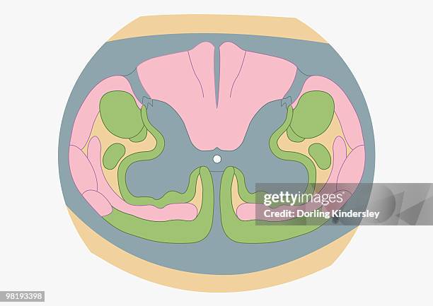
cross section digital illustration of spinal nerve fibres and convey motor signals highlighted in pink and green - motor neuron stock-grafiken, -clipart, -cartoons und -symbole
gray matter (spinal cord) with many multipolar motor neurons and unmyelinated nerve fibers, 50x - motor neuron stock-fotos und bilder
David Larkin from Dallas Crane performs to help raise funds for the fight against Motor Neuron Disease ahead of the 2018 Australian Open at Melbourne...
David Larkin from Dallas Crane performs to help raise funds for the fight against Motor Neuron Disease ahead of the 2018 Australian Open at Melbourne...
Jimmy Barnes performs at the MND concert to help raise funds for the fight against Motor Neuron Disease ahead of the 2018 Australian Open at...
Jimmy Barnes performs at the MND concert to help raise funds for the fight against Motor Neuron Disease ahead of the 2018 Australian Open at...
Jimmy Barnes performs at the MND concert to help raise funds for the fight against Motor Neuron Disease ahead of the 2018 Australian Open at...
Jimmy Barnes performs at the MND concert to help raise funds for the fight against Motor Neuron Disease ahead of the 2018 Australian Open at...
Jimmy Barnes performs at the MND concert to help raise funds for the fight against Motor Neuron Disease ahead of the 2018 Australian Open at...
Jimmy Barnes performs at the MND concert to help raise funds for the fight against Motor Neuron Disease ahead of the 2018 Australian Open at...
Jimmy Barnes performs at the MND concert to help raise funds for the fight against Motor Neuron Disease ahead of the 2018 Australian Open at...
Jimmy Barnes performs at the MND concert to help raise funds for the fight against Motor Neuron Disease ahead of the 2018 Australian Open at...
Jimmy Barnes performs at the MND concert to help raise funds for the fight against Motor Neuron Disease ahead of the 2018 Australian Open at...
Jimmy Barnes performs at the MND concert to help raise funds for the fight against Motor Neuron Disease ahead of the 2018 Australian Open at...
Jimmy Barnes performs at the MND concert to help raise funds for the fight against Motor Neuron Disease ahead of the 2018 Australian Open at...
Jimmy Barnes performs at the MND concert to help raise funds for the fight against Motor Neuron Disease ahead of the 2018 Australian Open at...
Jimmy Barnes performs at the MND concert to help raise funds for the fight against Motor Neuron Disease ahead of the 2018 Australian Open at...
Jimmy Barnes performs at the MND concert to help raise funds for the fight against Motor Neuron Disease ahead of the 2018 Australian Open at...