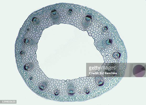
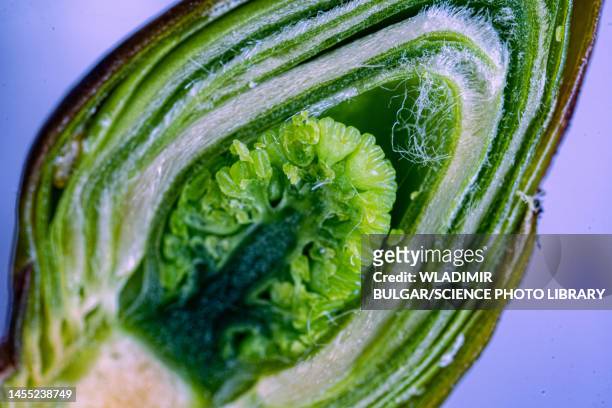
stem cross section. buttercup (ranunculus), herbaceous dicot, 8x. shows: vascular bundles arranged in a ring (typical of dicots), xylem, phloem, epidermis, cortex, and pith. - lichtmikroskopische aufnahme stock-fotos und bilder
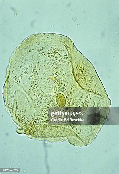
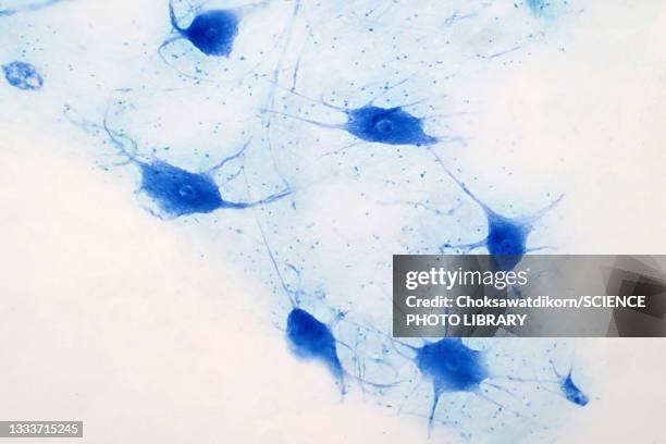
cheek cell. human squamous epithelial cell, mouth, 250x. shows: nucleus, cytoplastm and cell membrane. this is a very flat (or squamous) cell obtained inside the oral cavity. iodine stain. - lichtmikroskopische aufnahme stock-fotos und bilder
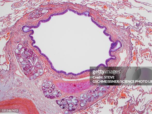
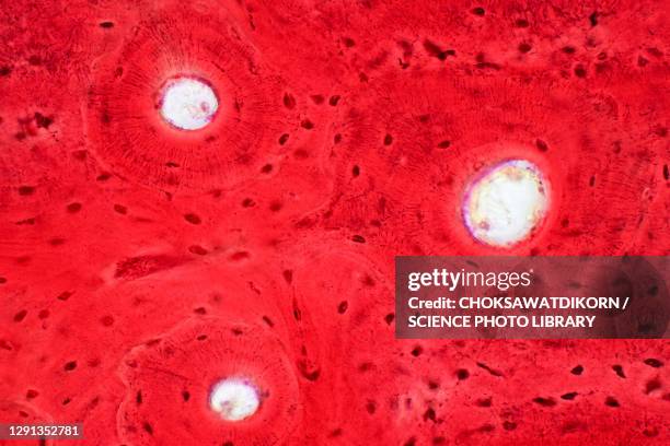
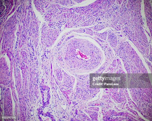
atherosclerosis of coronary artery, 5x at 35mm. reduced lumen. wall has excess calcification & fibrous connective tissue. h - lichtmikroskopische aufnahme stock-fotos und bilder
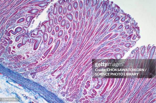
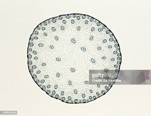
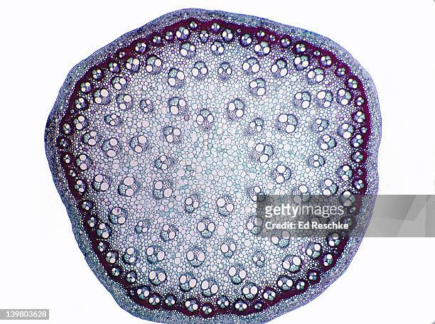
stem cross section. corn (zea), herbaceous monocot, 5x. shows: scattered vascular bundles typical of monocots, xylem, phloem, pith and epidermis. - lichtmikroskopische aufnahme stock-fotos und bilder
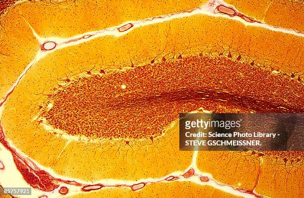
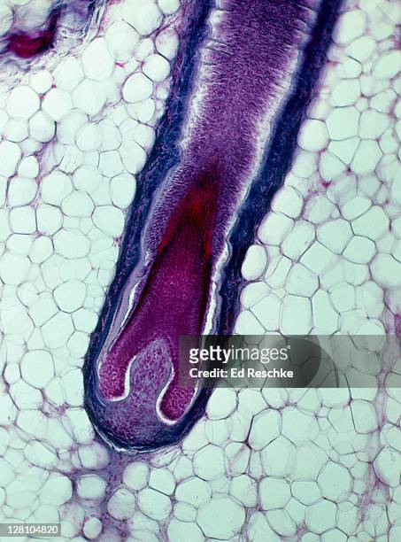
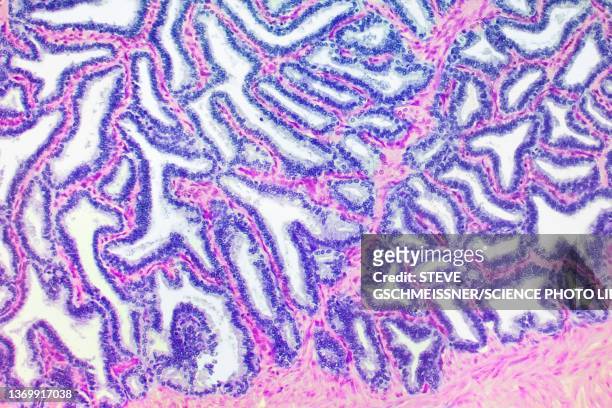
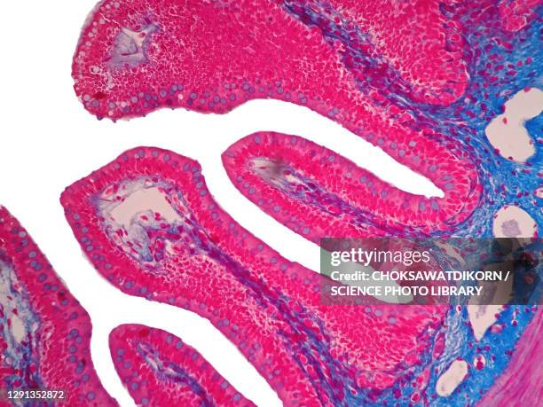
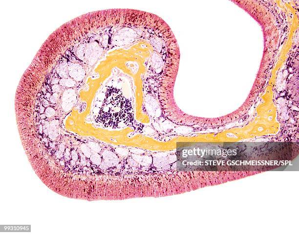
hair follicle, hair bulb, and hair papilla, human scalp, 25x at 35mm. growth takes place at the base of the hair follicle. adipose tissue surrounds the hair bulb. - lichtmikroskopische aufnahme stock-fotos und bilder
blood cells with a monocyte and platelets,100x light micrograph - lichtmikroskopische aufnahme stock-fotos und bilder
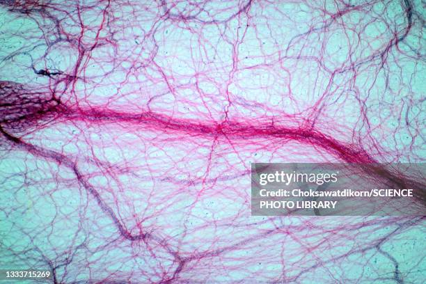
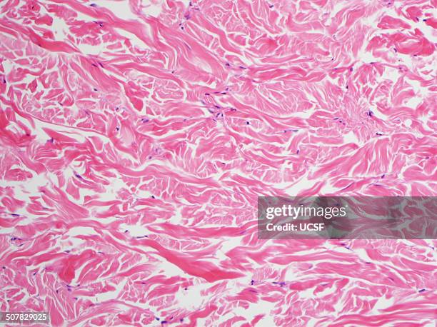
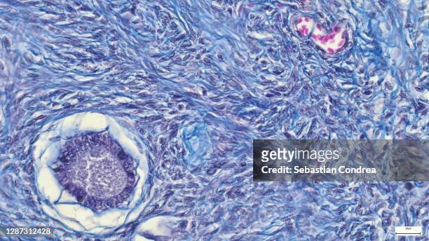
h&e stain, light microscopy, abundant collagen in a gardner fibroma - lichtmikroskopische aufnahme stock-fotos und bilder
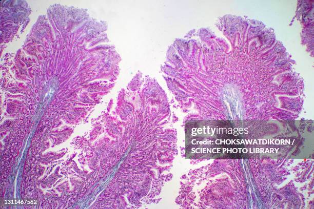
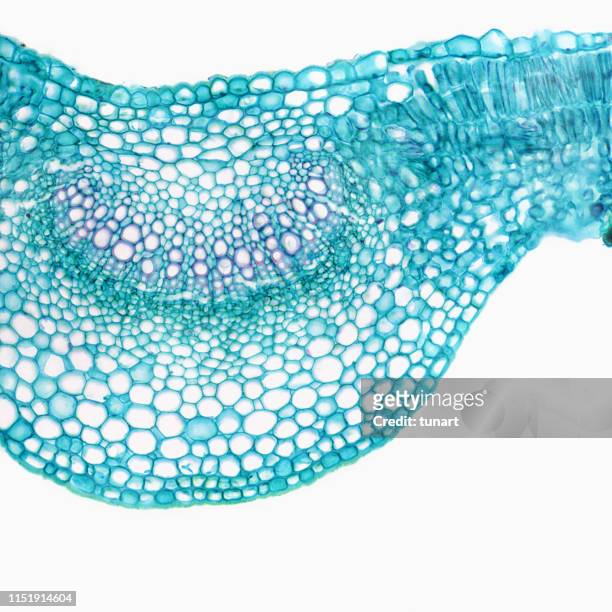
smilax, monocot stem cross section, 8x. shows: scattered vascular bundles, xylem, phloem, cortex and epidermis. - lichtmikroskopische aufnahme stock-fotos und bilder
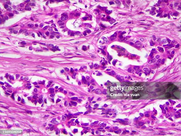
immunofluorescent photomicrograph, organs samples, histological examination, histopathology on the microscope - lichtmikroskopische aufnahme stock-fotos und bilder
alfalfa (medicago sativa) root nodosity cross section - lichtmikroskopische aufnahme stock-fotos und bilder
mikroskopische ansicht des kreuzes abschnitt des lilac-bleibes - lichtmikroskopische aufnahme stock-fotos und bilder
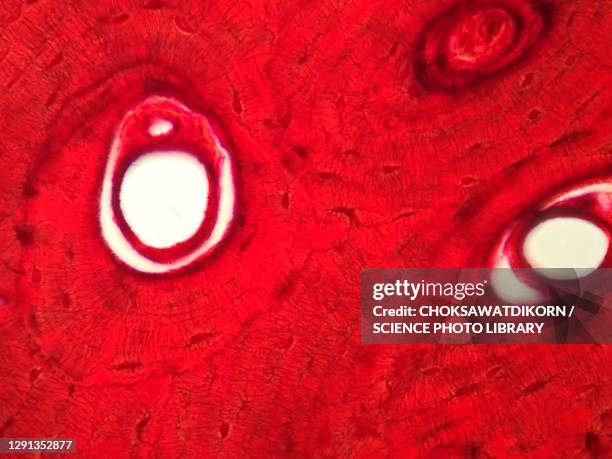
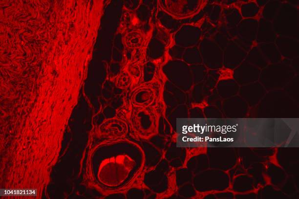
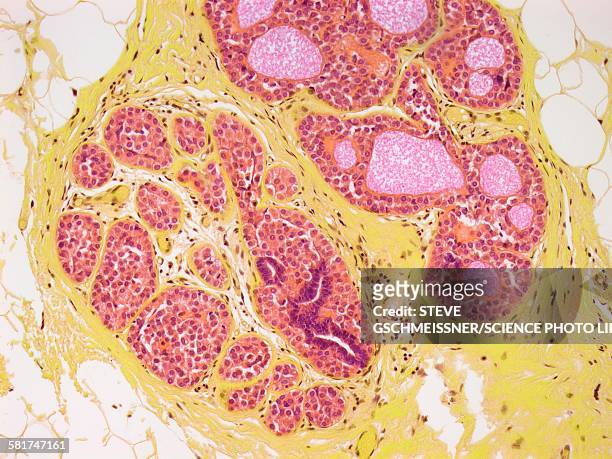
red fluorescence lipoma medical human tumour fatty tissue - lichtmikroskopische aufnahme stock-fotos und bilder
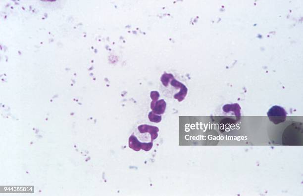
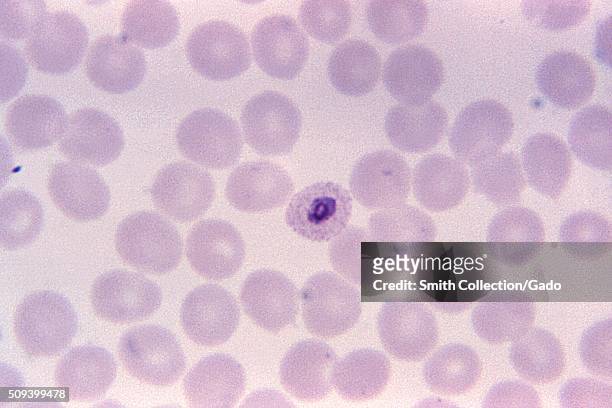
Photomicrograph of the malaria parasite Plasmodium Ovale growing as a double trophozoite in one red blood cell and a single trophozoite, on a thin...
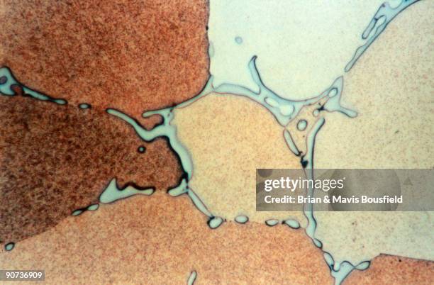
Aluminium 4% copper . Aluminium 4% copper . Grains of solid solution of copper in aluminium. Light micrograph in bright field. Magnification 500x.
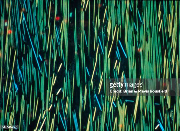
Carbon fibre in a polymer matrix. PMC carbon fibre in a polymer matrix. Light micrograph in crossed polar light. Magnification 100x.
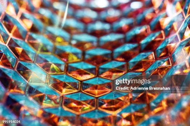
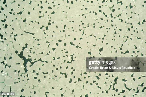
Glass reinforced plastic. Light micrograph. GRP glass reinforced plastic. Light micrograph in differential interference contrast. Magnification 100x.
Magnified 1125X, this thin-film, Giemsa-stained photomicrograph revealed the presence of a growing Plasmodium ovale trophozoite, with a ring nucleus....