menschliche innere organe vektorsymbole bearbeitbarer strich festlegen - human tissue stock-grafiken, -clipart, -cartoons und -symbole
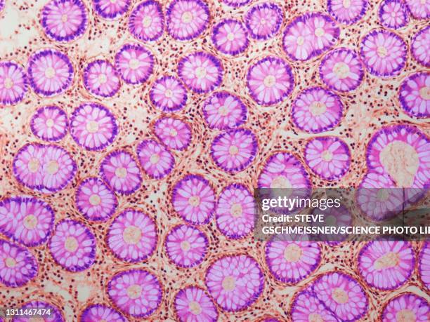
thymus. 10x shows: numerous lobules, cortex (darker with numerous lymphocytes), medulla (lighter). produces a hormone thymosin. - human tissue stock-fotos und bilder
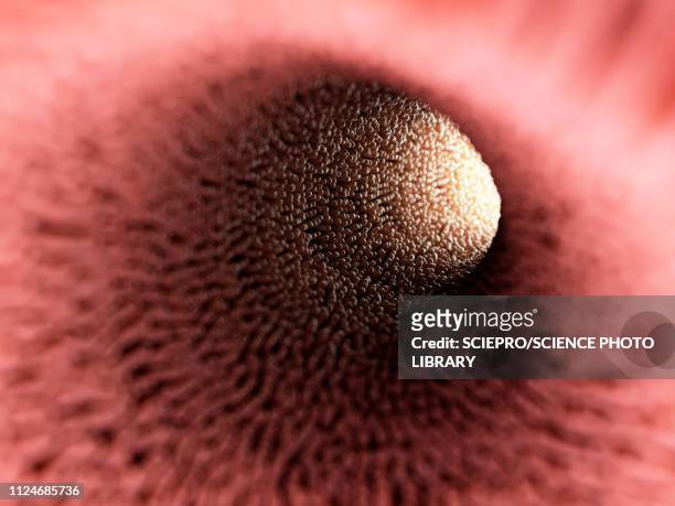
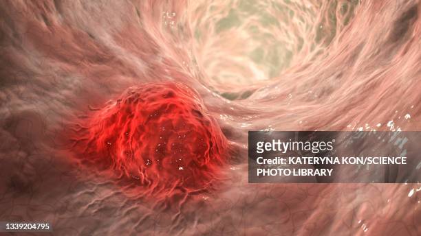
immunofluorescent photomicrograph, organs samples, histological examination, histopathology on the microscope - human tissue stock-fotos und bilder
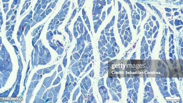
microscopy photography. cardiac muscle section, immunofluorescent photomicrograph, organs samples, histological examination, histopathology on the microscope. - human tissue stock-fotos und bilder
slide demonstrating breast tissue with ductal carcinoma. histopathology on the microscope. immunofluorescent photomicrograph, organs samples, histological examination, - human tissue stock-fotos und bilder
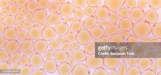
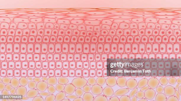
hautgewebezellen und fettgewebezellen, dermis und adipozyten - human tissue stock-grafiken, -clipart, -cartoons und -symbole
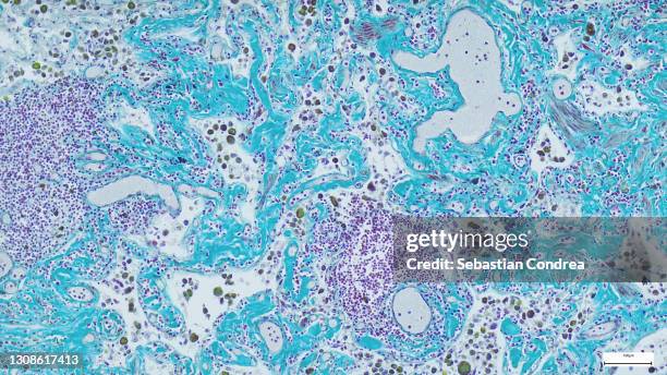
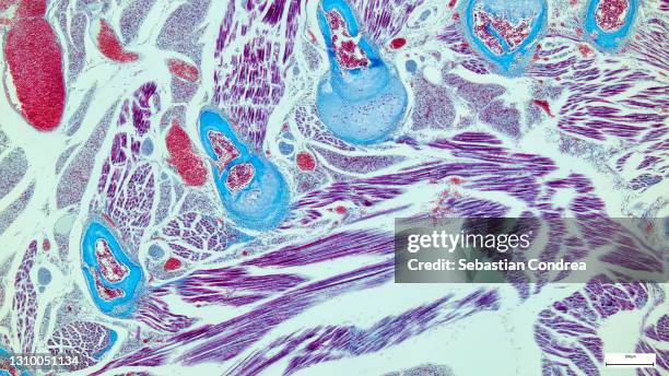
histological evaluation of the developing trabecular bone. goldner's trichrome staining with the blue/green colour representing mineralized tissue. immunofluorescent photomicrograph, organs samples. - human tissue stock-fotos und bilder
sampling strategy of the stem tissues. immunofluorescent photomicrograph, organs samples, histological examination, histopathology on the microscope - human tissue stock-fotos und bilder
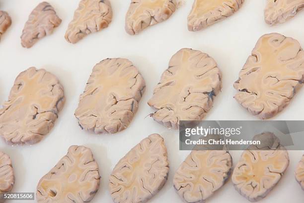
micrograph of rat brain. science cross section. immunofluorescent photomicrograph, organs samples, histological examination, histopathology on the microscope - human tissue stock-fotos und bilder
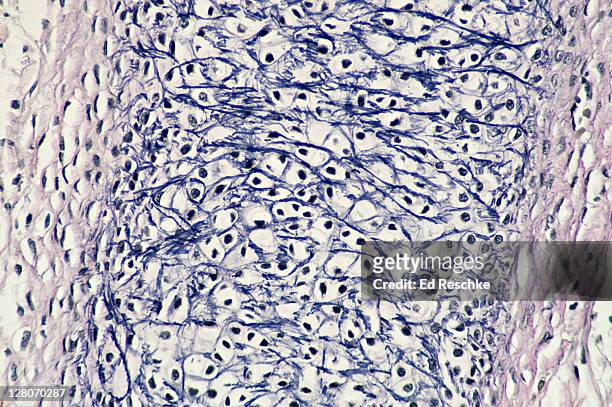
human fetal tissue. elastic cartilage; supporting connective tissue. (magnification x100). elastic fibres (dark) forming a network in the matrix, chondrocytes (cartilage cells) in lacunae. tissue has great tolerance for repeated bending. - human tissue stock-fotos und bilder
tissue in vascular plants that transports water and some nutrients. scientific research. plant tissue structure, immunofluorescent histopathology on the microscope - human tissue stock-fotos und bilder
education anatomy and physiology of tongue under the microscopic in laboratory. - human tissue stock-fotos und bilder
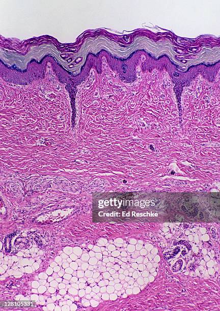
skin. epidermis, dermis, subcutaneous layer, thick skin, 25x at 35mm. shows: layers of epidermis, dermis, subcutaneous layer, sweat glands and ducts, adipose tissue. human. - human tissue stock-fotos und bilder
hautgewebezellen, hautschichten, blut in der vene - human tissue stock-grafiken, -clipart, -cartoons und -symbole
illustration of intestinal verticalilli - human tissue stock-grafiken, -clipart, -cartoons und -symbole
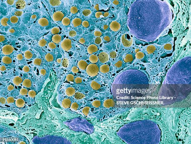
pancreas tissue, colored scanning electron micrograph - human tissue stock-grafiken, -clipart, -cartoons und -symbole
dense fibrous connective tissue; regular, tendon, 100x at 35mm. shows: layers of collagenous fibers (red), and rows of fibroblasts (dark) in parallel arrays. collagenous fibers and tendons have high tensile strength. - human tissue stock-fotos und bilder
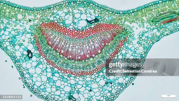
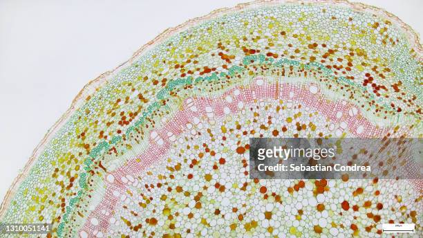
cross section - xylem is a type of tissue in vascular plants that transports water and some nutrients. scientific research. plant tissue structureimmunofluorescent photomicrograph, organs samples, histological examination, histopathology on the microscope - human tissue stock-fotos und bilder
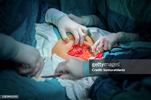
nuclear transfer being carried out on several embryonic stem cells used in cloning and genetic modification at laboratory - human tissue stock-fotos und bilder
hautgewebezellen, hautschichten, blut in der vene - human tissue stock-grafiken, -clipart, -cartoons und -symbole
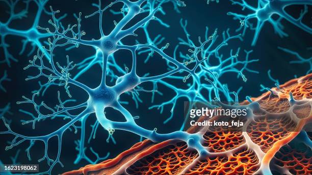
motor neuron (multipolar) with cell body and many processes (mostly dendrites), spinal cord (magnification x100). this multipolar motor neuron comes from the anterior (ventral) horn of the spinal cord grey matter. the stain is methylene blue and phloxine. - human tissue stock-fotos und bilder
cross-section plant stem under the microscope for classroom education. - human tissue stock-fotos und bilder
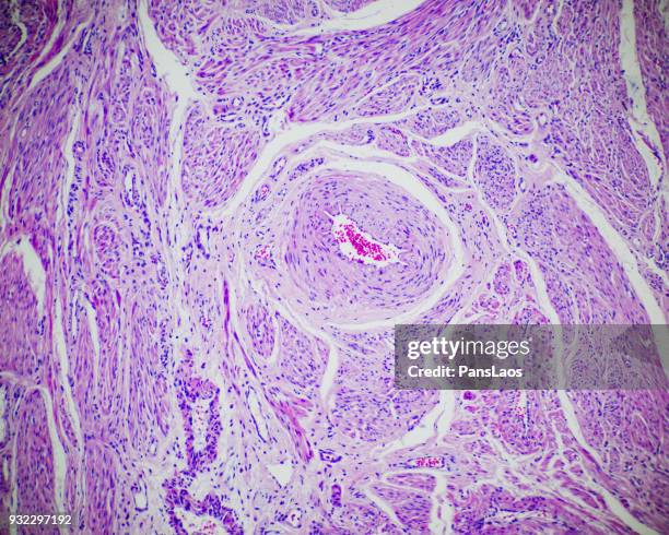
microscope of adenoid cystic carcinoma, rare type of cancer exist in many different body sites. this tumor occurs in the salivary glands, - human tissue stock-fotos und bilder
immunofluorescent photomicrograph,morphology and anatomy of the root system, organs samples, histological examination, histopathology on the microscope - human tissue stock-fotos und bilder
This photo taken on April 15, 2019 at the University of Tel Aviv shows a 3D print of heart with human tissue. Scientists in Israel on Monday unveiled...
carcinoma cell, colored transmission electron micrograph (tem) - human tissue stock-grafiken, -clipart, -cartoons und -symbole
menschlichen körper und die inneren organe - human tissue stock-grafiken, -clipart, -cartoons und -symbole