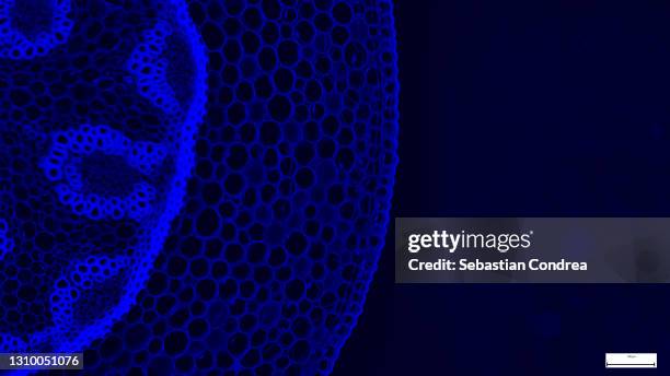
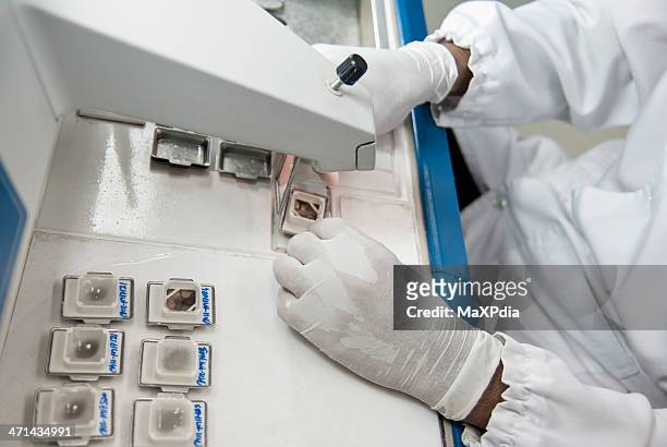
immunofluorescent photomicrograph, organs samples, histological examination, histopathology on the microscope - histology stock-fotos und bilder
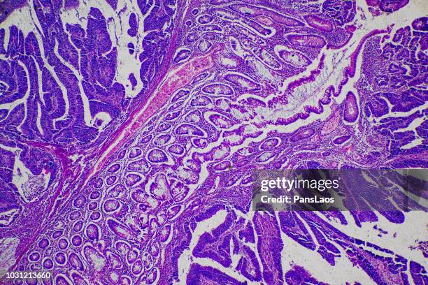
slide demonstrating breast tissue with ductal carcinoma. histopathology on the microscope. immunofluorescent photomicrograph, organs samples, histological examination, - histology stock-fotos und bilder
microscopic photo of a professionally prepared slide demonstrating breast tissue with ductal carcinoma. - histology stock-fotos und bilder
microscope of adenoid cystic carcinoma, rare type of cancer exist in many different body sites. this tumor occurs in the salivary glands, - histology stock-fotos und bilder
squamous epithelial cells of human cervix under the microscope view. pap smear test is a procedure to test for cervical cancer in women - histology stock-fotos und bilder
menschliches kompaktes knochengewebe, struktur von knochengewebe oder knochengewebe, osteon - histology stock-grafiken, -clipart, -cartoons und -symbole
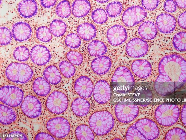
pancreas. serous acini, immunofluorescent photomicrograph, organs samples, histological examination, histopathology on the microscope. - histology stock-fotos und bilder
microscopy photography. cardiac muscle section, immunofluorescent photomicrograph, organs samples, histological examination, histopathology on the microscope. - histology stock-fotos und bilder
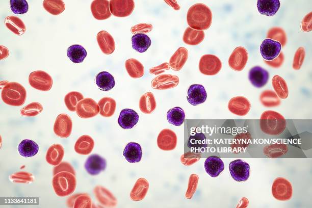
acute lymphoblastic leukaemia, illustration - histology stock-grafiken, -clipart, -cartoons und -symbole
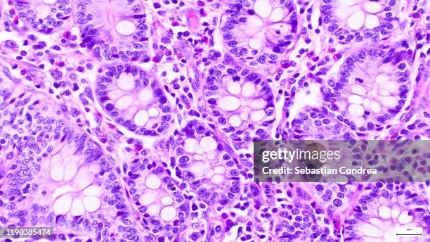
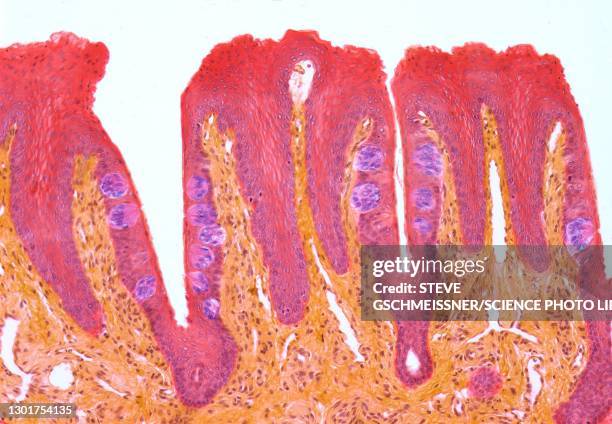
villi in the small intestine (ileum, human), simple columnar epithelium and intestinal glands, 50x - histology stock-fotos und bilder
small foramen magnum meningioma seen after gadolinium injection on axial t1 mri image - histology stock-fotos und bilder
histological evaluation of the developing trabecular bone. goldner's trichrome staining with the blue/green colour representing mineralized tissue. immunofluorescent photomicrograph, organs samples. - histology stock-fotos und bilder
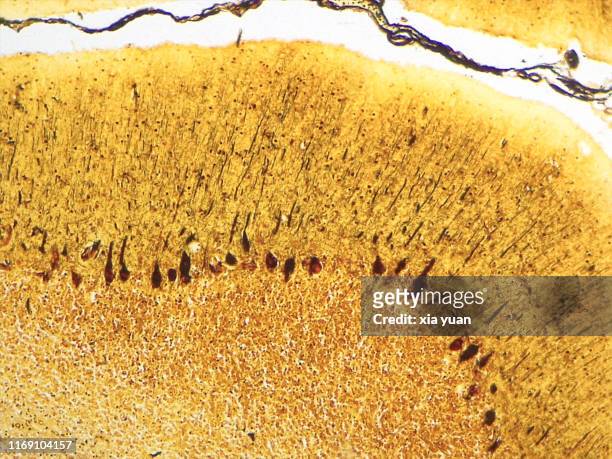
cross section of the cerebellum and nerve human under the microscope for education in lab - histology stock-fotos und bilder
pyramidal neurons in the cerebral cortex, illustration - histology stock-grafiken, -clipart, -cartoons und -symbole
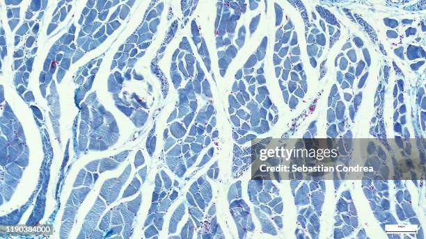
histology (or microscopic anatomy) lining a bronchus, pseudostratified ciliated columnar epithelium, 100x - histology stock-fotos und bilder
oesophageal cancer, illustration and micrograph - histology stock-grafiken, -clipart, -cartoons und -symbole
oesophageal cancer, illustration and micrograph - histology stock-grafiken, -clipart, -cartoons und -symbole
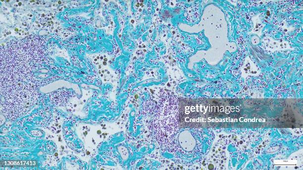
immunofluorescent photomicrograph,microscopic photograph of a professionally prepared slide of liver tissue. the liver is divided histologically into lobules. organs samples, histological examination, histopathology on the microscope - histology stock-fotos und bilder
micrograph of rat brain. science cross section. immunofluorescent photomicrograph, organs samples, histological examination, histopathology on the microscope - histology stock-fotos und bilder